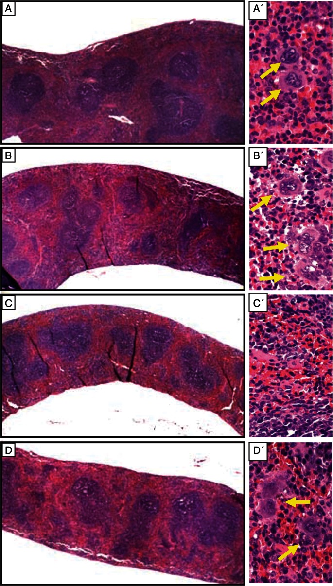
Fig. 5.
Histological sections of spleens from mice exposed to irradiation, with or without treatment with MGN-3, at 1 week post-irradiation. Cross-sections (A–D left panel) of the spleens and higher magnifications of each (A′–D′ right panel). A) Control group, no exposure to irradiation or treatment with MGN-3. B) Mice treated with MGN-3 alone. C) Mice exposed to irradiation alone. D) Mice treated with MGN-3 and exposed to irradiation. Please notice the yellow arrows pointing to the megakaryocytes.

