Fig. 2.
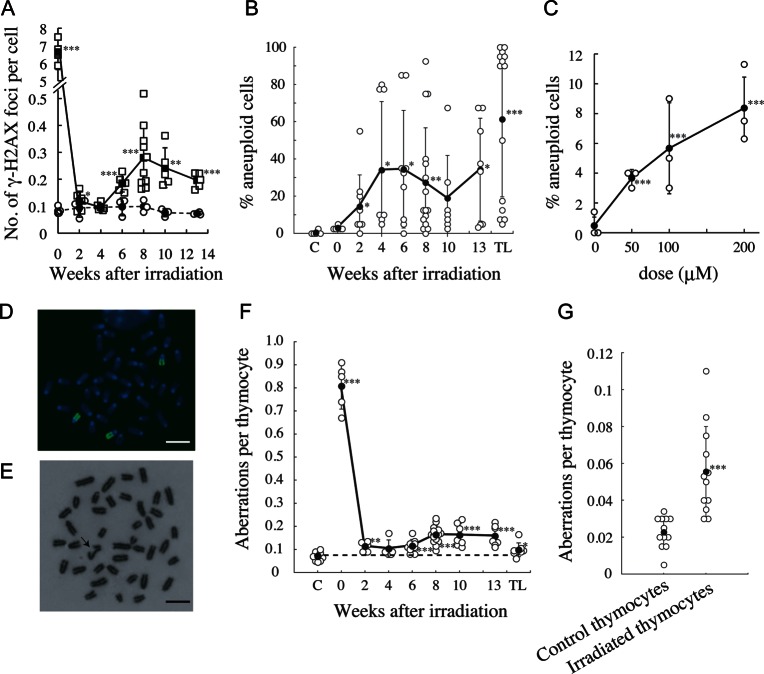
Delayed induction of γ-H2AX foci and chromosomal instability in post-irradiation thymocytes. Symbols are the same as those in Fig. 1 except that in (A), open squares and closed squares indicate individual irradiated mouse data and mean values ± standard deviations, respectively. Open circles and closed circles indicate individual control mouse data and mean values ± standard deviations, respectively. TL, thymic lymphoma. (A) Delayed induction of γ-H2AX foci after irradiation. (B) Delayed appearance of aneuploidy after irradiation. (C) Induction of aneuploidy by H2O2 in thymocytes. (D) Trisomy 15 in a thymocyte 8 weeks after irradiation. Bar, 10 μm. (E) Chromatid break (arrow) in a thymocyte 8 weeks after irradiation. Bar, 10 μm. (F) Delayed induction of chromosomal aberrations in post-irradiation thymocytes cultured with proliferative stimuli for 48 h. (G) Induction of chromosomal aberrations in vivo in thymocytes 8 weeks after irradiation.

