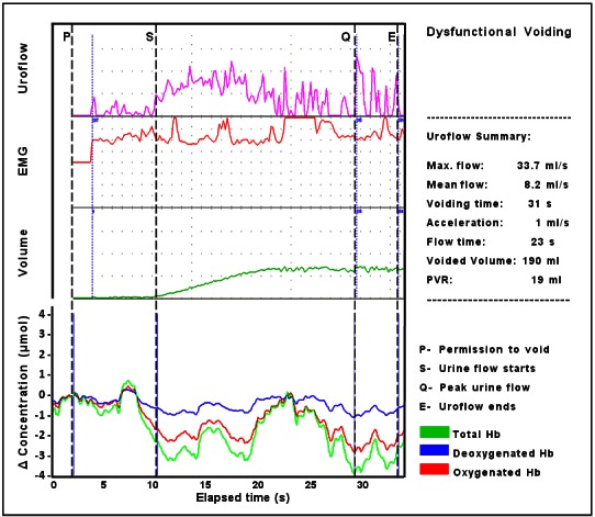
Fig. 4.

Simultaneous data (uroflow, EMG, voided volume, and NIRS pattern of chromophore change in a child with symptoms of non-neurogenic lower urinary tract dysfunction. Following permission to void an initial increase in ΔtHb occurs due to an equal increase of ΔO2Hb and ΔHHb; a sharp decrease in ΔtHB is then evident predominantly due to a fall in ΔO2Hb that continues as uroflow starts, periodic increases in ΔtHB/ΔO2Hb occur, but the trend is negative during voiding particularly from 23 seconds, while ΔHHb remains stable. This chromophore pattern implies a failure of oxygen supply to meet demand following permission to void followed by hemodynamic fluctuation with variations in the availability of O2Hb and total blood volume in the detrusor during voiding.
