
Figure 4. Physical half-life of trans-spliced vs non-trans-spliced test mRNA in Ascaris embryos.
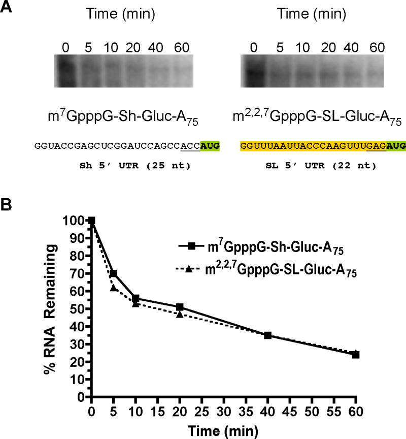
A). Decay of labeled Gaussia luciferase RNAs in embryos. 32P-labeled RNAs were introduced into embryos (see Suppl. Figure 1 for additional RNA information; orange shaded sequence is the SL), RNA extracted at intervals, the RNA resolved by electrophoresis, and visualized by phosphoimager analysis. B). Decay curves of 32P-labeled Gaussia RNAs in A. RNA levels illustrated in A were quantitated by phosphoimager analysis and plotted using levels observed at time zero as 100%. Similar data were obtained using transfection of unlabeled RNAs and analysis by real-time PCR (data not shown).