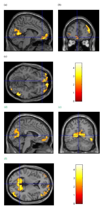
Figure 1.

Group grey matter volume effects. Cluster in the frontal lobes of significantly lower grey matter volume in cocaine-dependent subjects relative to healthy controls. Cursor location: X=−6.0, Y=43.5, Z=−12.0, left medial orbitofrontal cortex (a and c). Also shown: left cuneus and precuneus region (a) in the posterior cortical cluster, and right dorsolateral prefrontal cortex (b). d–f: Cluster of significantly lower grey matter volume in cocaine-dependent subjects relative to healthy controls that includes the precuneus and cuneus regions surrounding the parietal-occipital sulcus. Cursor location: X=−3.0, Y=−58.5, Z = 6.0. Colour bar indicates voxel-wise t-statistics
