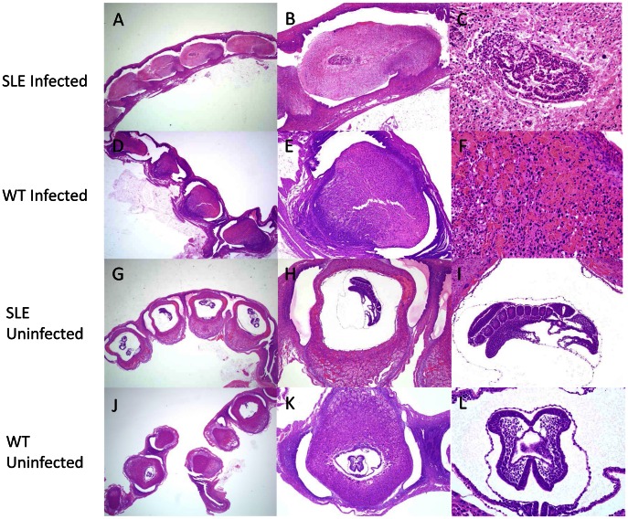
Figure 3. Malaria caused massive decidual necrosis in SLEs and control animals.
Uterine section of an infected SLE mouse (A) or infected WT mouse (D) showing massive decidual necrosis at day 8 post-conception at low-magnification (1.25×). High-magnification views (4× and 10×) of uteri from an infected SLE (B–C) and infected WT mouse (E–F). Uterine section of an uninfected SLE (G) or uninfected WT mouse (J) showing normal morphology of an 8 day old fetus at low-magnification (1.25×). High-magnification views (4× and 10×) of uteri from a non-infected SLE (H–I) or uninfected WT mouse (K–L). Uteri from all mice were analyzed by a pathologist (n = 72).

