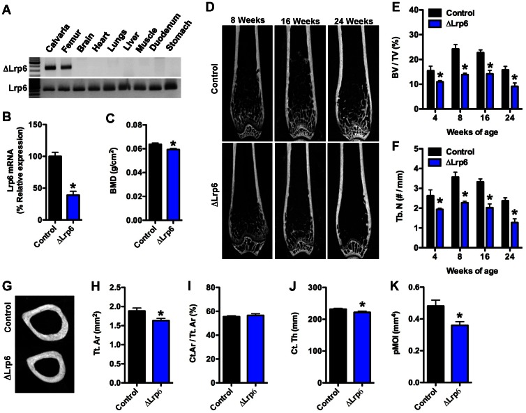
Figure 2. Trabecular and cortical bone acquisition is reduced in ΔLrp6 mice.
(A) PCR analysis of Cre-mediated recombination of the Lrp6flox allele in ΔLrp6 mice. (B) qPCR analysis of Lrp6 mRNA levels in the femur of control and ΔLrp6 mice. (C) Whole-bone mineral density assessed by DXA at 6months of age (n = 16–26 mice). (D) Representative microCT images of bone structure in the distal femur of control and ΔLrp6 mice at 8, 16, and 24 weeks of age. (E) Quantification of trabecular bone volume per tissue volume (BV/TV) in the distal femur. (F) Quantification of trabecular numbers (Tb.N) in the distal femur. (G) Representative microCT images of cortical bone structure at the femoral mid-diaphysis in 24 weeks old control and ΔLrp6 mice. (H) Cortical tissue area (Tt.Ar). (I) Cortical bone area per tissue area (Ct.Ar/Tt.Ar). (J) Cortical thickness (Ct. Th). (K) Polar moment of inertia (pMOI). For microCT analyses, n = 5–8mice/genotype. *p<0.05.

