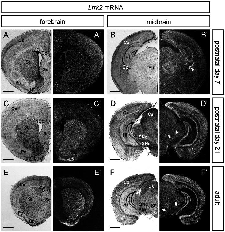
Figure 2. Expression analysis of Lrrk2 mRNA in the forebrain and midbrain of postnatal mice.
ISH for Lrrk2 mRNA in coronal sections of forebrain (left part) and midbrain (right part) from postnatal day 7, postnatal day 21 and adult mice. Note that expression of Lrrk2 is highly dynamic in the postnatal forebrain. While the expression level in the striatum and the olfactory tubercle seem to increase dramatically during development, the Lrrk2 level in cortex remain rather unchanged (A,C,E). On the level of the midbrain, ISH signals for Lrrk2 augment considerably in the hippocampus and cortex, while the level in midbrain structures like the Substantia nigra pars compacta (white arrows, SNc) remain quite low (B,D,F). Abbreviations: Cc, corpus callosum; Cx, cortex; Cp, choroid plexus (white arrowhead in B’); Cs, superior colliculus; Hi, hippocampus; Ot, olfactory tubercle; Pc, piriform cortex; Pn, parafascicular nucleus; Rn, red nucleus; Se, septum; SNc, SN pars compacta; SNr, SN pars reticulata; St, striatum. Scale bars represent 1 mm.

