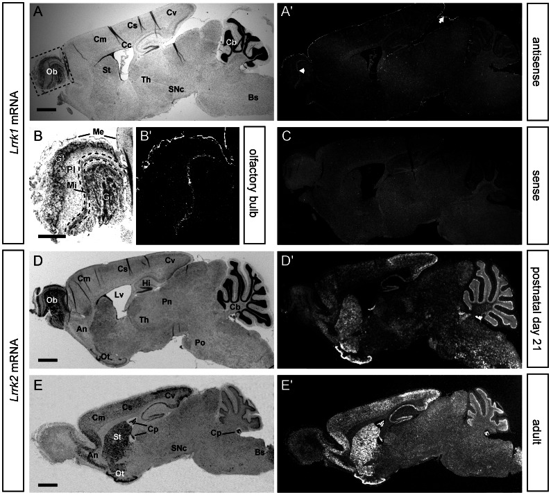
Figure 3. Comparative expression analysis of Lrrk1 and Lrrk2 mRNA in the brain of adult mice.
ISH for Lrrk1 (top part) and Lrrk2 (bottom part) mRNA in sections from P21 (A–D) and adult mice (E). Note that Lrrk1 mRNA is barely detectable in the adult mouse brain and only visible in the non-neuronal meninges (white arrow) and the olfactory bulb (white arrowhead) (A). A detailed view onto the adult olfactory bulb depicts the solely neuronal expression of Lrrk1 in the mitral cell layer (B). Specificity of the Lrrk1 signals were verified by using the corresponding sence-probe as negative control (C). In contrast, strong Lrrk2 expression can be detected in various regions throughout the postnatal (D) and adult CNS (E). Abbreviations: An, anterior olfactory nucleus; Bs, brain stem; Cb, cerebellum; Cc, corpus callosum; Cm, motor cortex; Co, cortex; Cp, choroid plexus; Cs, somatosensory cortex; Cv, visual cortex; Gl, glomerular layer; Gr, granual layer; Hi, hippocampus; Me, meninges; Mi, mitral layer; Ob, olfactory bulb; Ot, olfactory tubercle; Pn, parafascicular nucleus; Pl, plexiform layer; Po, pons; SNc, substantia nigra pars compacta; St, striatum; Th, thalamus; Lv, lateral ventricle. Scale bars represent 1 mm (A, D, E) and 500 µm (B).

