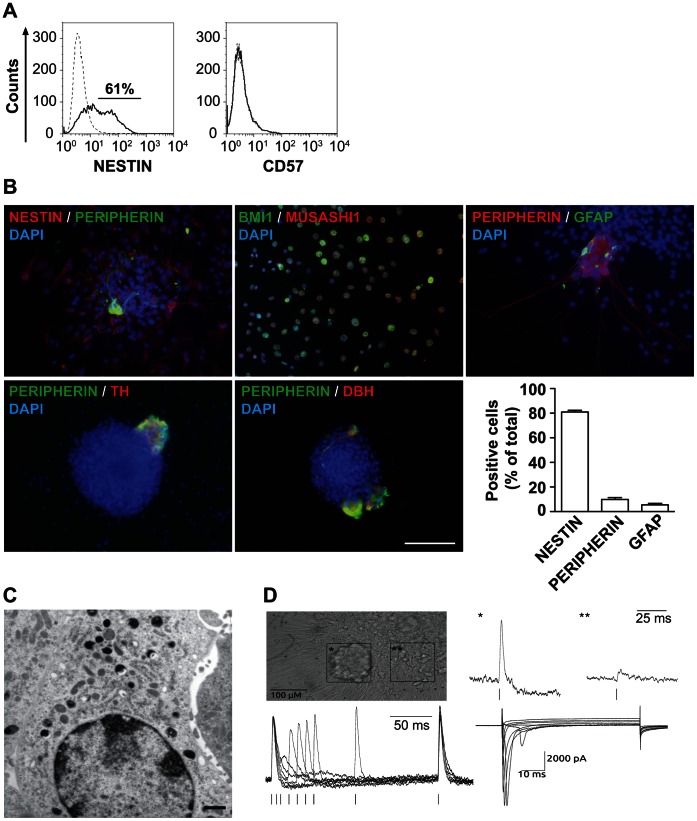
Figure 5. Adrenal spheres contain many SAP-like cells and few sympathetic neurons and glial cells.
(A) A majority of sphere cells express nestin, while none express CD57. Flow cytometric analysis of dissociated adrenal-derived sphere cells stained for nestin (left panel) and CD57 (right panel). Dashed lines define isotype controls and solid lines specific antigen expression. (B) The majority of adrenal-derived sphere cells expresses nestin, BMI1 and MUSASHI1 while few cells express peripherin or GFAP. Immunofluorescence microscopy of spheres allowed to attach overnight on poly-D-lysine/fibronectin-coated glass coverslips in 1% FCS-containing sphere medium, analyzed for co-expression of nestin, peripherin, BMI1, MUSASHI1, GFAP, TH, DBH. Nuclei were counterstained with DAPI. Scale bar equals 100 µm. Quantification of positive cells is shown in the histogram. (C) Adrenal-derived spheres contain a minority of cells with dense core vesicles. Representative electron microscopic image of a cell from an adrenal-derived sphere. Scale bar equals 1 µm. (D) Adrenal-derived spheres harbor many immature appearing cells lacking functional voltage-gated channels and few mature appearing neurons exhibiting electrical activity. Microphotograph showing a neuronal-like cell cluster (*) and an area outside of the cluster (**). Fluorescence change (ΔF) in di-8-ANEPPS stained preparations corresponding to compound action potentials (CAP) from the cluster (*) and lack of CAP in the area outside of it (**), upper right panel. Representative traces of double pulse stimulation, whereby the second CAP was elicited at Δt of 100, 50, 40, 30, 20, 10, and 5 ms, lower left panel. Representative voltage-activated whole-cell inward currents recorded from a cell depolarized in 5 mV steps from –70 to +35 mV for 60 ms, from a holding potential of −120 mV, lower right panel.

