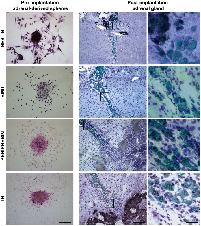
Figure 7. Adrenal-derived sphere cells integrate in situ where they downregulate expression of progenitor markers but do not differentiate to chromaffin cells.
Pre-implantation adrenal-derived spheres attached to glass coverslips (left column) and 3 week post-implantation rat adrenal gland prior implanted with CFSE-labeled sphere cells (middle and right columns) were stained for nestin, BMI1, peripherin and TH. Stainings were visualized by immunohistochemistry. In the middle and right columns, fluorescent images (CFSE, green) are overlaid on immunohistochemistry images. Images in the right column are magnifications of images shown in the middle column. Scale bars are 200 µm (left and middle columns) and 50 µm (right column).

