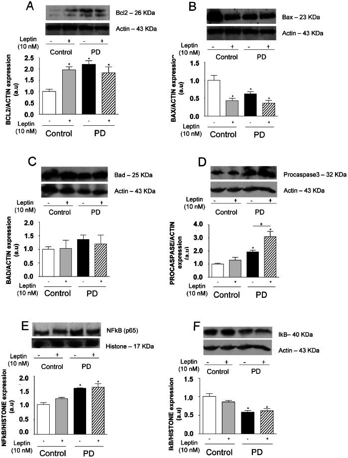
Figure 6. Thymocytes from PD rats present alterations in the pro- and anti-apoptotic protein levels and NF-kB nuclear translocation in basal condition.
Thymocytes isolated from control and PD rats were incubated in the presence or absence of leptin (10 nM) for 24 hours, and BcL-2, Bad, Bax, procaspase 3, NF-kB, IkB, actin and histone protein expression were assessed in total (BcL-2, Bad, Bax, procaspase3, IkB and actin) or nuclear (NF-kB and histone) extracts by western blotting analysis. Quantification of bands is expressed in arbitrary units. Values are means ± S.E of 6 animals/group. *P<0.05 compared to control group.

