
Figure 7.

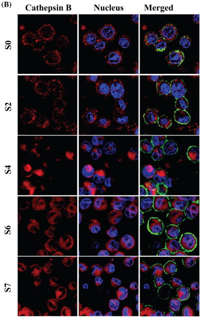
Comparison of the effects of CeO2 nanorods with different aspect ratios on cathepsin B release from damaged lysosomes and the initiation of NALP 3 inflammasome activation in THP-1 cells. THP-1 cells were seeded into 8-well chamber slides and incubated with CeO2 nanorods of different aspect ratio at 80 μg mL−1 for 4 h in complete RPMI 1640. After fixation and permeabilization, cells were stained with Magic Red™ (ImmunoChemistry Technologies), wheat germ agglutinin 633 and Hoechst 33342 dye, followed by visualization under a confocal 1P/FCS inverted microscope as described in the Material and Method section. Monosodium urate (MSU) crystals were used as the positive control. (A) The control cells exhibited intact punctuate lysosomes stained by the red fluorescent cathepsin B substrate, while MSU treated cells showed a diffuse red color in the cytoplasm as a result of lysosome damage. (B) In agreement with the cellular data in Figure 5, S0 and S2 materials had no effect on cathepsin B release compared to prominent lysosome damage by the higher aspect ratio materials (S4, S6 and S7).