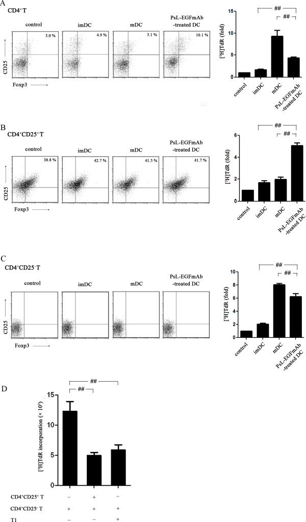
Figure 5.
Allogeneic mixed T cell proliferation assays. imDCs, mDCs or PsL-EGFmAb-treated mDCs were co-cultured with freshly isolated human CD4+, CD4+CD25+ and CD4+CD25– T cells, respectively, then proliferation assays were performed and the results are shown as fold increases in [3H]TdR incorporation. Flow cytometric analysis was also performed to detect CD4+CD25+Foxp3+ Tregs. A, DCs co-cultured with CD4+ T cells. B, DCs co-cultured with CD4+CD25+ T cells. C, DCs co-cultured with CD4+CD25– T cells. D, A suppression assay was performed to evaluate the suppressive function of CD4+CD25+ T cells using the same method. Tregs were either co-cultured with PsL-EGFmAb-treated mDCs (T1) or freshly isolated from health adults. The mean ± SD of four independent experiments is shown. #p < 0.05, ##p < 0.01.

