
Figure 3.
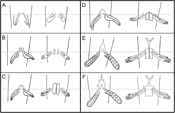
Schematic representation of pelvic development in the Australian lungfish and the axolotl. Each panel compares pelvic development of the Australian lungfish (left side of each panel) and the axolotl (right side). The grey line marks the position of the acetabulum. A) Beginning of pelvic development through cartilaginous condensations at the acetabula. B) The condensations extend anteriorly for the lungfish (pubis) and posteriorly (ischium) and slightly anteriorly (pubis) for the axolotl. C) The pubis of the lungfish continues to extend anteriorly and the pubis of the axolotl grows anteriorly. D) The pubis of the lungfish is now fused at the midline and is triangular shaped. The axolotl pubis is complete anteriorly and the ischium is complete posteriorly. E) Anterior growth of the lungfish pelvic process and appearance of the ypsiloid cartilage in the axolotl. F) Adult morphology of the pelvic girdle in both species. Anterior is at the top.