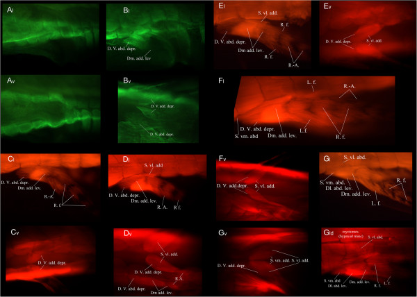
Figure 4.
Pelvic musculature development in the Australian lungfish. Immunostained larvae of Neoceratodus forsteri showing the developing pelvic musculature. All stages were incubated in a primary antibody against skeletal muscle. A and B were visualized through a secondary anti-mouse 488 Alexa antibody and C and D were visualized with a secondary anti- IgG1(γ1) 568 Alexa antibody. v: ventral view and l: lateral view. A) Stage 50, B) Stage 51, C) Stage 52, D) Stage 54, E) Stage ‘56’, F) Stage ‘61’, G) Stage ‘63’. Dl. Abd. lev., dorsolateral abductor levator; Dm. add. lev., dorsomesial adductor levator; D. V. abd. depr., deep ventral abductor depressor; D. V. add. depr., deep ventral adductor depressor; L.f., Lepidotrichia flexors; R.-A., radial-axials; R. f., radial flexors; S. vl. abd., superficial ventrolateral abductor; S. vl. add., superficial ventrolateral adductor; S. vm. abd., superficial ventromesial abductor. Anterior to the left.

