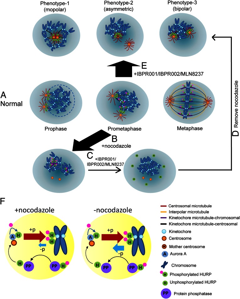
Fig. 6.
Models for nucleation of centrosomal and kinetochore microtubule fibers by Aurora A-regulated HURP phosphorylation during spindle formation. (A) As a cell enters mitosis, the nuclear envelope (light blue circle) breaks down; HURP is expressed and associated with the minus end of centrosomal microtubules that project toward chromosomes. As the cell cycle proceeds to prometaphase and metaphase, HURP gradually is phosphorylated and translocated to the vicinity of chromosomes to assist nucleation and stabilization of kinetochore fibers. Finally, bipolarity is established. Phosphorylated HURP forms a rod-like structure (purple bar) that links to kinetochore. (B) Treatment with 300–400 nM nocodazole enriched the phosphorylated HURP that nucleates kinetochore microtubules. (C) Adding IBPR001/IBPR002/MLN8237 disrupts nucleation of the kinetochore microtubule. (D) Removing nocodazole under the treatment with IBPR001/IBPR002/MLN8237 reinitiates tubulin polymerization from centrosomes but not from kinetochores. HURP goes to the minus end of centrosomal microtubules that face toward the chromosomes. (E) Inhibiting HURP phosphorylation by IBPR001/IBPR002/MLN8237 abolishes nucleation of HURP in the vicinity of chromosomes. The unphosphorylated HURP distributes restrictively to the minus end of centrosomal microtubules. Because IBPR001/IBPR002/MLN8237 do not completely block the separation of the duplicated centrosomes, HURP is preferentially associated with the microtubules that emanate from the mother centrosome, which resides in proximity to the chromosomes. (F) Models for HURP phosphorylation and dephosphorylation with and without nocodazole. In the presence of nocodazole, the force that drives HURP phosphorylation (by Aurora A) overrides dephosphorylation (through a PP1/PP2A-dependent pathway). Conversely, the ratio of phosphorylation/dephosphorylation is decreased in the absence of nocodazole.

