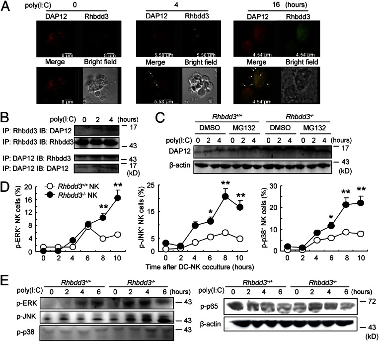
Fig. 4.
Inhibition of TLR3-triggered NK cell DAP12 expression and MAPK activation by Rhbdd3. (A) Splenocytes were stimulated with poly(I:C) for the indicated time, and then NK cells were isolated. The intracellular localization of Rhbdd3 (red) and DAP12 (green) were determined by confocal analysis. (Objective, 40×; numerical aperture, 1.4.) Bar lengths are as indicated. (B) Cell lysates from poly(I:C)-activated splenic NK cells were immunoprecipitated and immunoblotted by anti-Rhbdd3 antibody or anti-DAP12 antibody as indicated. (C) Rhbdd3+/+ or Rhbdd3−/− splenic NK cells were treated with MG132 (20 μM) or DMSO (vehicle control) for 2 h and then stimulated by poly(I:C) for the indicated time. The expression of DAP12 was analyzed by Western blotting. (D) Rhbdd3+/+ or Rhbdd3−/− splenic NK cells were stimulated with poly(I:C) in the presence of BMDCs. The levels of p-ERK, p-JNK, and p-p38 in NK1.1+ NK cells were determined using a Phosflow method (BD Biosciences). (E) Splenic NK cells were, respectively, purified from Rhbdd3+/+ and Rhbdd3−/− mice i.p. treated with poly(I:C) and D-GalN. The expression of p-ERK, p-JNK, p-p38, and p-p65 were evaluated by Western blotting. The data shown are the means ± SD (D) from three independent experiments. *P < 0.05; **P < 0.01; NS, not significant.

