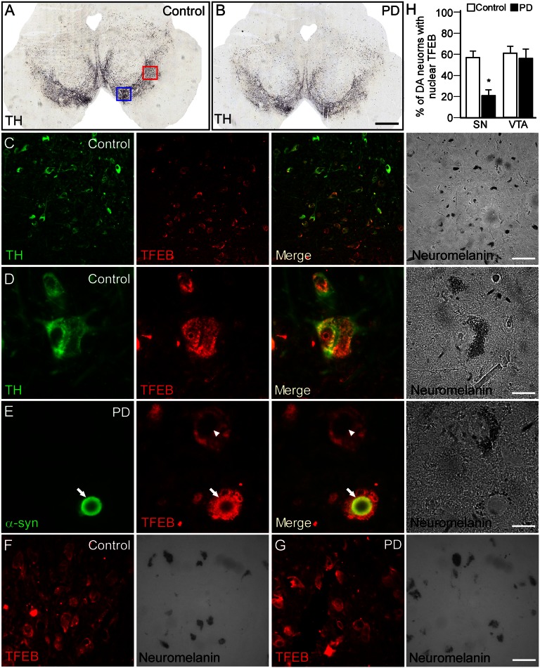
Fig. 2.
Changes in TFEB localization in human PD brains. (A and B) TH immunohistochemistry showing the DA neurons in control and PD human midbrain. Note the reduced density of TH+ neurons in the PD brain consistent with the clinical diagnostic. Red and blue squares in A indicate the regions analyzed to study the expression of TFEB in nigral (shown in C–E) and VTA neurons (shown in F and G), respectively. (Scale bar, 500 μm.) (C and D) Double-immunofluorescence staining of human postmortem midbrain showed that TFEB is well expressed in neuromelanin-containing nigral DA neurons (C). (Scale bar, 60 μm.) High-power magnification revealed that TFEB is localized both in the cytoplasmic and nuclear compartments of A9 neurons (D). (Scale bar, 12 μm.). (E) In PD brains, although the expression level did not seem to be altered, TFEB was not observed in the nuclear compartment and appeared clustered in the cytoplasm (arrowhead). In addition, TFEB colocalized with α-syn in Lewy bodies containing nigral neurons (arrow). Identity of the DA neurons was further confirmed by the expression of neuromelanin (light microscopy). (Scale bar, 12 μm.). Immunofluorescence staining of TFEB from control (F) and PD (G) human midbrain showing that its pattern of expression is unchanged in VTA DA neurons. (Scale bars, 30 μm.) (H) Percentage of nigral and VTA DA neurons presenting TFEB immunoreactivity in the nucleus. A total of 200 neuromelanin-containing cells from five different brains were examined in both the VTA and SN in control and PD human brains.

