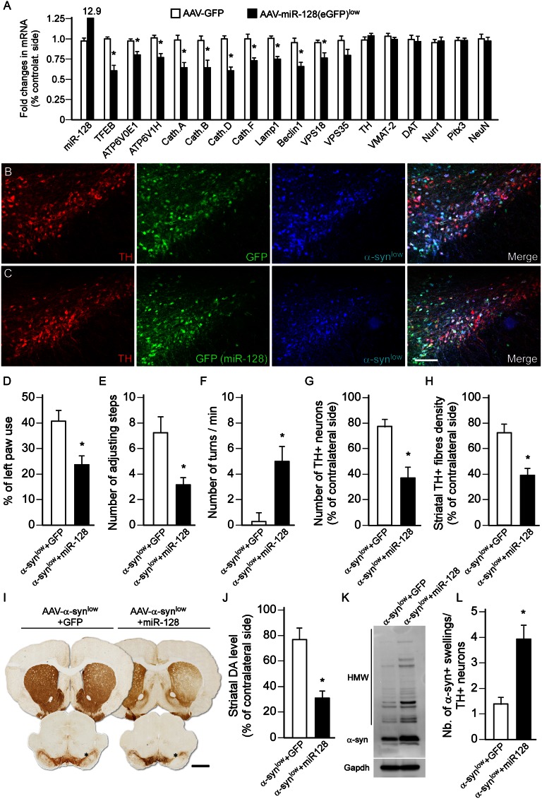
Fig. 4.
miR-128-induced inhibition of autophagy aggravates α-syn toxicity. (A) Midbrain expression levels of ALP and dopaminergic markers, analyzed by qPCR 3 wk after intranigral injection of AAV-GFP (open bars) or AAV–miR-128 (eGFP) (low titer, 1.2 × 10¹⁰ gc/mL, black bars). *P < 0.05 compared with GFP group (Student t test; n = 5 per group). (B and C) Triple-immunofluorescence staining showing the colocalization of TH (red), GFP (green in B), miR-128 (green, visualized by the GFP reporter in C, low titer 1.2 × 10¹⁰ gc/mL), and α-syn (low titer: 3.8 × 10¹¹ gc/mL, blue) in nigral DA neurons 3 wk after vector injection. (Scale bar, 100 μm.) (D–F) Motor function was assessed at 8 wk in rats overexpressing α-syn at low titer together with GFP or miR-128 using the cylinder test (D), stepping test (E), and amphetamine-induced rotation test (F). miR-128 overexpression (low titer 1.2 × 10¹⁰ gc/mL) together with α-syn promoted the development of behavioral deficits on the side controlateral to the vector injection in these three tests compared with the GFP-overexpressing animals. *P < 0.05 compared with α-syn+GFP group (Student t test; n = 8 per group). (G–I) Brain sections were stained for TH (I) and survival of nigral DA neurons (G) and loss of striatal innervation (H) was determined at 8 wk by stereological counting and optical densitometry, respectively. Measurements showed that miR-128 overexpression at low titer aggravated α-syn toxicity (low titer: 3.8 × 10¹¹ gc/mL) and triggered a significant loss of nigral TH+ neurons and striatal TH+ terminals compared with GFP-overexpressing rats. *P < 0.05 compared with α-syn+GFP group (Student t test; n = 8 per group). (Scale bar, 1.5 mm.) Asterisk indicates the injection site. (J) HPLC measurement showed that striatal DA concentration was significantly lower in rats coexpressing α-syn+miR-128 compared with the GFP controls. *P < 0.05 compared with α-syn+GFP group (Student t test; n = 5 per group). (K) Western blot analysis showed that miR-128–mediated inhibition of autophagy enhanced the accumulation of HMW striatal α-syn oligomers (n = 5 per group, both vectors delivered at low titer). (L) The load of α-syn was estimated by calculating the ratio of the number of striatal α-syn accumulated axonal swellings over the number of surviving nigral TH+ neurons (G). miR-128 overexpression (low titer) significantly increased the burden of α-syn aggregation in these structures. *P < 0.05 compared with α-syn+GFP group (Student t test; n = 8 per group). Data in J–L were all obtained at 8-wk survival.

