Fig. 5.
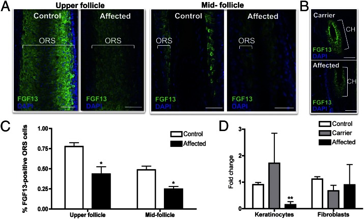
Immunofluorescence staining reveals that FGF13 expression is dramatically reduced in affected hair follicles compared with control. (A) Immunofluorescence staining in control and affected anagen hair follicles reveals a decrease in FGF13 localization throughout the outer root sheath (ORS) in the mid and upper portions of the hair follicle. (B) Immunofluorescence staining in carrier and affected telogen hair follicles reveals decreased FGF13 expression in the affected hair follicle, recapitulating the dosage effect seen at the mRNA level. Z-stack images were taken using identical settings and a consistent Z-stack interval between control, carrier, and affected samples. CH, club hair of a telogen follicle. (C) Quantification of the percentage of FGF13-expressing ORS cells in control and affected hair follicles reveals a decrease in the number of FGF13-expressing cells within the upper and midfollicle regions of the ORS (P < 0.05). Data represent the averaged value of three independent experiments, where images taken at a 40× magnification were used to quantify the number of FGF13-positive cells relative to the total number of ORS cells. For immunofluorescence studies, hair follicles were stained from three control and two affected skin biopsies. (D) qRT-PCR revealed that FGF13 expression is reduced in keratinocytes but not in fibroblasts grown from skin biopsies. A Student t test was performed with a cutoff P value of 0.05 for statistical significance; *P < 0.05, **P < 0.01. Error bars represent the SEM. (Scale bar, 100 μm.)

