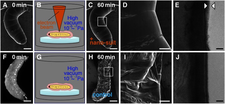
Fig. 1.
(A–D) A living Drosophila larva was exposed to high vacuum with electron-beam irradiation for 60 min. (F and G) Before SEM observation, a different larva (light micrograph in F) was placed in the observation chamber without electron-beam irradiation for 60 min. (H and I) The specimen collapsed completely when subsequently observed by SEM. Each small white square in C and H is shown magnified in D and I, respectively. (E and J) TEM images are shown of vertical sections through the surface of each animal. The layer between the arrowheads in E indicates the limits of the newly formed outer membrane, not present in J. An outer layer covering the animal represents ECSs in B and G. [Scale bars: 0.3 mm (A, C, F, and H), 0.1 mm (D and I), and 0.2 µm (E and J).]

