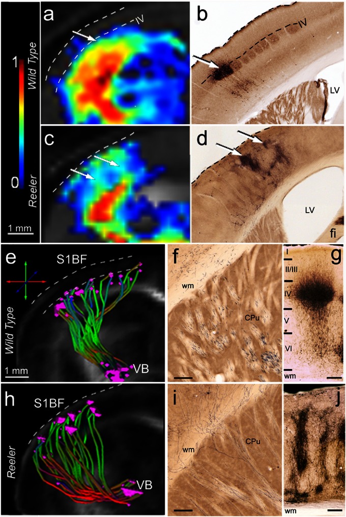
Fig. 5.
Intracortical in vivo diffusion-based fiber mapping (A, C, E, and H) and histological tracing (B, D, F, G, I, and J) show remodeling of the thalamocortical terminal field in reeler mutant compared with wild-type mice. (A–D) Probability maps of connectivity (A and C) depicting fine details of the intracortical portion of the wild-type (A) and reeler (C) TCP, similar to the staining pattern seen in the cortical areas of the same mice after MiR tracer injections (B and D). Note the distribution of the termination field of the wild-type pathways (A and B) at the level of cortical layer IV and the extended columnar representation in reeler animals (C and D, arrows). (E and H) Selective 3D reconstruction of wild-type (E) and reeler (H) thalamic fibers originating in the VB (MiR injection site) and reaching S1BF. The reconstructions reveal subcortical defasciculation and oblique intracortical trajectories of the reeler somatosensory thalamocortical projections (H) compared with wild-type mice (E). The origin and the target points of the identified pathway were plotted for each fiber. The target points of the reeler trajectories are distributed across the whole cortical thickness (H), contrasting to their clustering at the level of cortical layer IV (upper border depicted by lower dashed line) in wild-type mice (E). (F–J) Representative subcortical (F and I) and intracortical (G and J) MiR tracing of the wild-type (F and G) and reeler (I and J) TCP. Note the pattern of reeler subcortical axons (I), reaching the cortex in oblique trajectories, very similar to the pathway identified with global fiber tracking. In S1BF, the main terminal field of the wild-type thalamic projections (G) is layer IV. The reeler axons are spanning nearly the entire cortical thickness, forming a peculiar columnar pattern (J). (Scale bars, 250 µm.)

