Fig. 4.
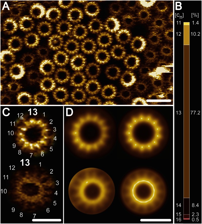
High-resolution AFM topographs of reconstituted B. pseudofirmus OF4 extWT c-rings. An isolated extWT B. pseudofirmus OF4 c13 ring sample was densely reconstituted in lipid vesicles and high-resolution topographic images were recorded in quasi 2D-crystalline regions. (A) Overview. Higher-protruding (lighter) and lower-protruding (darker) rings become visible, showing the cytoplasmic and periplasmic ends of the extWT c-rings, respectively (20, 23, 27). No central lipid plugs are visible (28). (B) extWT c-ring stoichiometry statistics ranging from c11 to c16 (n = 270), with a majority of 77.2% c13 stoichiometry. [cn] represents the stoichiometry, and [%] is the percentage of the c-rings. (C) Unprocessed images of cytoplasmic (Upper) and periplasmic (Lower) sides with countable 13 subunits. (D) Reference-free single-particle averages of the cytoplasmic (Upper; n = 350) and periplasmic (Lower; n = 390) sides of the c13 rings. Nonsymmetrized (Left) and symmetrized (Right) averages confirm 13-fold symmetry of the complex. The average diameter of the cytoplasmic side of the c13 ring is 6.7 ± 0.3 nm (n = 30). (Scale bars: 10 nm in A; 5 nm in C and D.)

