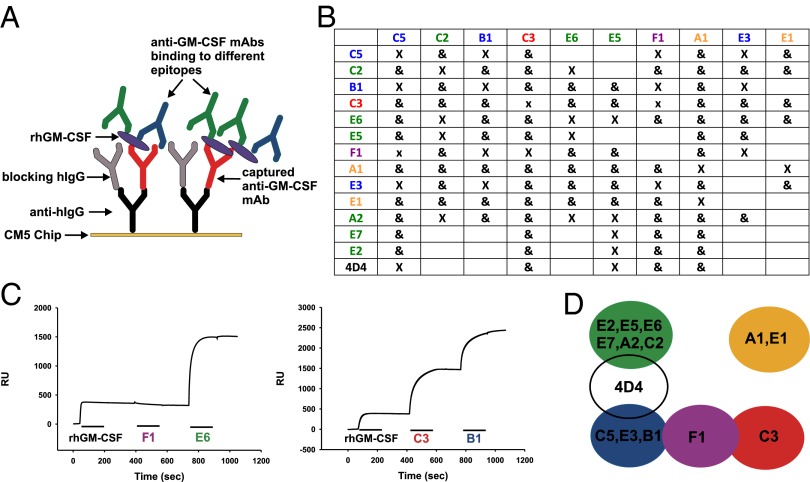
Fig. 2.
Human mAbs against GM-CSF target multiple epitopes. SPR was used to map the binding of anti–GM-CSF mAbs to rhGM-CSF. (A) A schematic showing how an anti-hIgG is immobilized on a CM5 sensor chip to capture the first anti–GM-CSF mAb injected. After blocking of free sites with an excess of hIgG, rhGM-CSF is injected and bound by the first mAb. Subsequently, a second and third anti–GM-CSF mAb is injected, and the binding is monitored in real time by SPR. (B) A summary of the epitope-mapping experiments, with “X” meaning that the two mAbs cannot bind simultaneously and “&” meaning they can bind simultaneously to rhGM-CSF. mAbs in the leftmost column denote the captured mAb for each experiment. (C) Representative mapping experiments. (Left) After capture of C3 and binding of rhGM-CSF, only E6, and not F1, can bind. (Right) After capture of A1 and binding of rhGM-CSF, both C3 and B1 can bind simultaneously. (D) A map of the GM-CSF epitopes. Color coding is maintained throughout. The 4D4 is a mouse mAb raised against rhGM-CSF.

