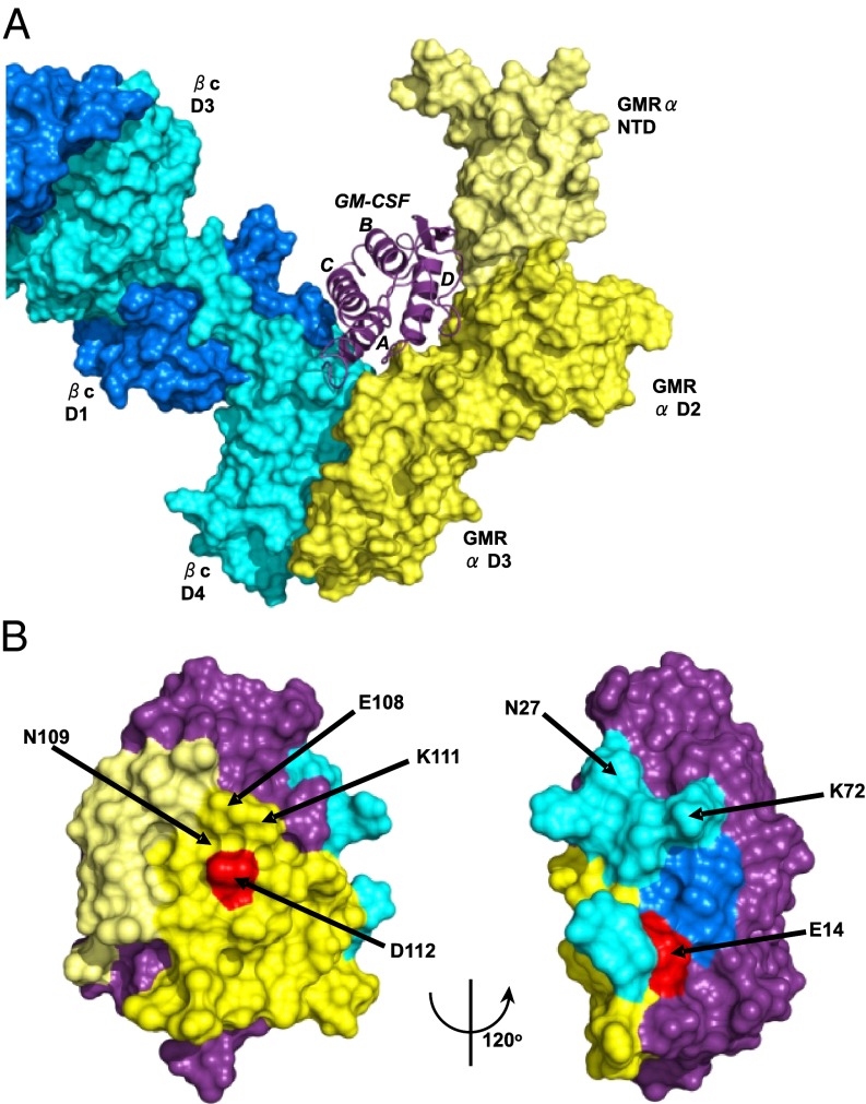
Fig. 3.
Mutations on the surface of GM-CSF and area buried by the subunits of the GM-CSF receptor. (A) The GM-CSF:receptor ternary model. GM-CSF is shown in purple ribbon, the GMR-α in yellow molecular surface (with the NTD shown in lighter shade), and the βc shown in blue (different shades for the two monomer chains of the βc dimer). (B) Two views of the surface of GM-CSF using the coloring scheme in A. The surfaces that interact with the GMR-α are in yellow, with the GMR-βc in blue and the remainder of the cytokine colored purple. Some of the residues targeted for mutation are highlighted, and key residues, shown by mutation to markedly disrupt antibody binding, are shown in red.

