Fig. 2.
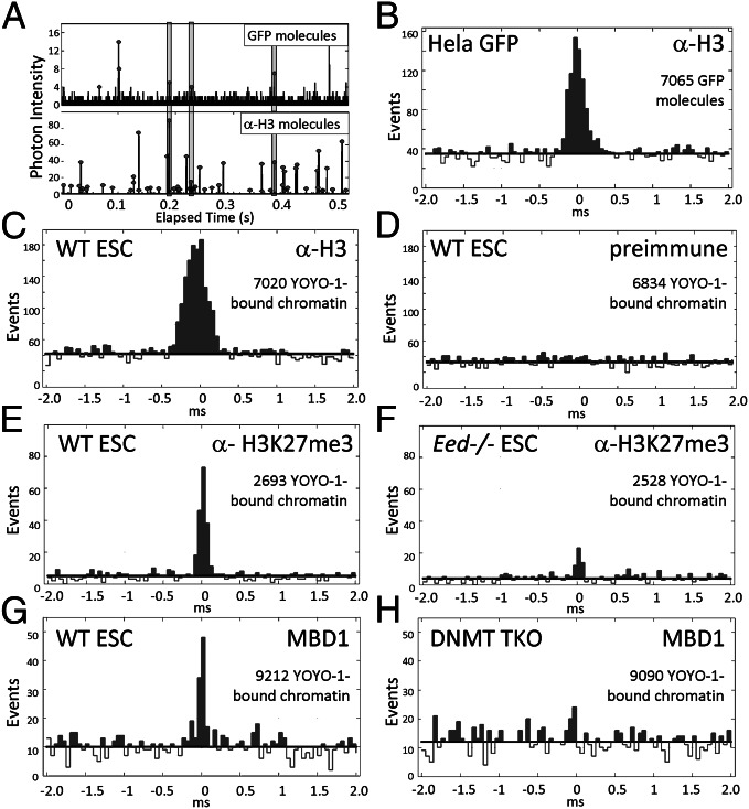
SCAN detects chromatin features with high specificity. AlexaFluor dye-tagged antibodies or MBD1 protein were bound to chromatin before analysis by SCAN. (A and B) Chromatin was from H2B-GFP expressing HeLa cells, and antibody (α-H3) was H3-specific. (A) Time-resolved photon counts (0.5 s) reporting GFP (Upper) and α-H3 (Lower) fluorescent emissions, depicted as peaks. Shading identifies antibody-bound chromatin complexes emitting both fluorophores nearly simultaneously. (B) Time-offset histogram for 7,065 GFP chromatin molecules analyzed in A. GFP fluorescent events are placed at time 0 and the time offset identifies how soon before or after the GFP emission AlexaFluor dye was detected. The peak centered at time 0 with width less than the transit time for molecules passing through the inspection volume (∼1 ms) identifies antibody–chromatin complexes. See Fig. S2 for description of coincidence calculations. (C and D) Chromatin was from WT-ESC bound to α-H3 (C), or preimmune mouse serum (D). (E and F) Antibody probe was specific for H3K27me3 and chromatin was from WT-ESC (E) or Eed−/− ESC, which are deficient for H3K27me3 (F). (G and H) MBD1 probe was specific for mC in duplex DNA, and chromatin was from WT-ESC (G) or DNMT TKO ESC, which are deficient for mC (H). Chromatin in C–F was detected by labeling with the intercalator YOYO-1, whose emission defined the 0 time in the offset plots.

