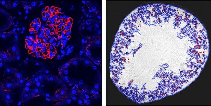
Fig. 4.

Intravenously injected CF is detected with fluorescence immunohistochemistry and MRI in low levels in the proximal tubule of a perfused kidney. A: red immunofluorescence of ferritin is seen in a glomerulus and surrounding tubules. B: image processing is used to detect CF labeling of the glomeruli (red dots) and apparent tubules in the surrounding cortex (blue lines) based on the intensity of the MRI voxels. (From Bennett KM and colleagues, unpublished observations).
