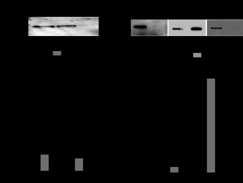
Fig. 4.
Distribution of VNUT60 in Calu-3 cells. A: postmitochondrial supernatant (S3, see Fig. 2) was centrifuged at 150,000 g for 3 h, and the resulting pellet (P4) and supernatant (S4) were analyzed by Western blot. Values [percentage of recovery relative to postnuclear supernatant (S1)] are means ± SD from ≥3 independent experiments. B: plasma membrane-rich fraction was isolated using the Plasma Membrane Protein Extraction kit. Equivalent aliquots of the resulting plasma membrane and cellular organelle membrane (Org) fractions were analyzed by Western blotting (VNUT) and slot blotting (MUC1 and GM130) and quantified as described in Fig. 2 legend. Values are means ± SD from ≥3 independent experiments.

