
Figure 5. Vascular phenotypes in low-dose foxC1dMO embryos.
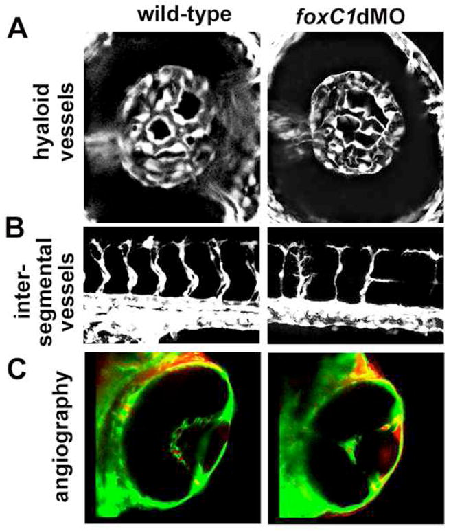
A. Hyaloid vasculature as revealed by fli1a:GFP in wild-type (left) and in low-dose foxC1dMO morphant embryos (right) at 48hpf. No differences in overall morphology was noted in low-dose morphants. C. Intersegmental vessels in wild-type (left) and low-dose foxC1dMO morphant (right) embryos at 48 hpf. D. Microangiography in wild-type (left) and low-dose foxC1dMO morphant embryos (right) at 4 dpf. Note the increased leakage of dye into the anterior chamber of foxC1dMO eyes.