
Figure 3.
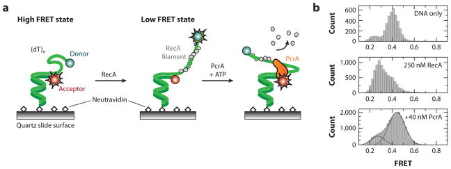
(a) A smFRET-based assay for observing the RecA-clearing activity of PcrA helicase. A DNA oligonucleotide with a Cy3-Cy5 FRET dye pair is affixed to the surface of a passivated microscope slide. Entropically driven collapse of the ssDNA brings the two dyes within efficient FRET distance. Addition of RecA leads to a rigid nucleoprotein filament, which separates the two dyes and reduces the observed FRET signal. In the presence of ATP, PcrA helicase clears RecA and reels the free DNA ends closer together. (b) smFRET data. (Top) Naked ssDNA brings both dyes relatively close, leading to a FRET signal of ~0.5. (Middle) Addition of RecA elongates the ssDNA tail and reduces the smFRET to ~0.3. (Bottom) In the presence of ATP, PcrA clears RecA from ssDNA as it reels the two DNA ends close to each other.