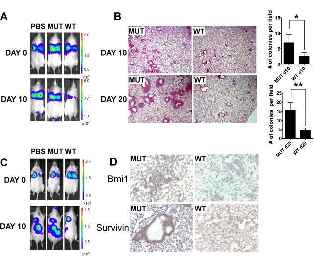
Figure 7. ARF 26-44 peptide blocks colonization of intravenously inoculated p53 null tumors.
A, ICR SCID mice were intravenously inoculated with CreERT2 Foxm1 fl/fl and p53 −/− sarcoma cells. Luciferase intensity was monitored with IVIS image machine following peptide treatment at 10 days after initial injection and right after injection at day 0. B, H&E staining of the lung tissue section from MUT or WT peptide treated mice at day 10 and day 20 after initial sarcoma cell injection and quantification of the number of the colonies per field of the corresponding lung tissue section. C, ICR SCID mice were intravenously inoculated with CreERT2 Foxm1 fl/fl and p53 −/− thymic lymphoma cells. Luciferase intensity was monitored with IVIS image machine following peptide treatment at 10 days after initial injection and right after injection at day 0. D, Representative pictures of Bmi1 and Survivin IHC staining of colonized sarcoma cells in the lung after either MUT or WT peptide treatment at day 20.

