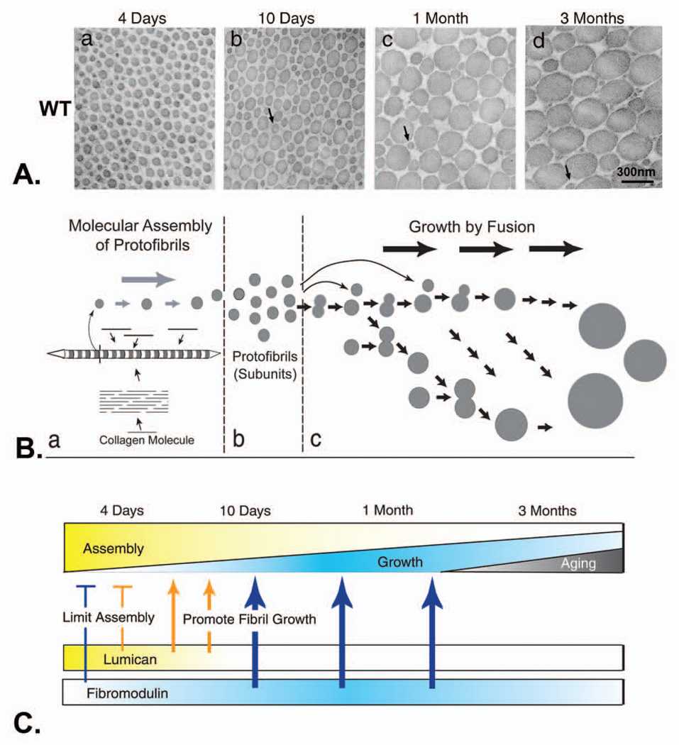
Fig. 4. Lumican and fibromodulin cooperatively regulate collagen fibrillogenesis in developing mouse tendon.
A. Collagen fibril structure during development in wild-type mouse tendons. Transmission electron micrographs of transverse sections from wild type mouse flexor tendons (a–d). Fibril structure was analyzed at different developmental stages, 4 days (a), 10 days (b), 1 month (c), and 3 months (d) postnatal. Bar, 300 nm. Arrows indicate protofibrils. B. Model for regulation of fibril growth by lumican and fibromodulin. The steps in collagen fibrillogenesis during tendon development are presented (a–c). (a) In the early steps of fibril formation, the molecular assembly of collagen monomers into protofibrils occurs in the pericellular space. Collagen molecules (bars) assemble into quarter-staggered arrays forming protofibrils, seen here as striated structures with tapered ends in longitudinal section and in cross section as circular profiles. Growth in length and diameter is by accretion of collagen at this stage. (b) Protofibrils in mouse tendons are stabilized through their interactions with fibril-associated macromolecules such as SLRPs. (c) Fusion of the protofibrils generates the mature fibril in a multistep manner. Progression through this growth process could be both by additive fusion (i.e., protofibrils add to its product, indicated by horizontal arrows) and by like-fusion (i.e., products from different steps can only fuse with like products, indicated by arrows in the oblique direction). C. The proportion of the assembly and growth steps occurring during development is illustrated. During the early stages, assembly is the main event and its relative contribution gradually decreases to the minimum degree necessary for maintenance at maturation. The relative contribution of growth by fusion increases to ~1 month. This is followed by a decrease during maturation (bottom). The expression data for lumican and fibromodulin is illustrated. The phenotypes observed in mutant mice indicate stage-specific regulatory mechanisms. At 4 days, both lumican and fibromodulin limit the assembly of the collagen monomers (bars). Characteristic of 10 days, growth from preformed protofibrils begins, and changes in both lumican and fibromodulin promote the transition from assembly to fibril growth by fusion (thin arrows). At later stages, only fibromodulin promotes the growth steps (thick arrows) Modified from Ezura et al. [127]

