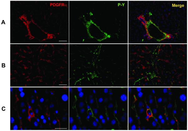
Fig. 4.
Localization of PDGFRα in the normal liver. PDGFRα and phospho-tyrosine (P-Y) double immunostaining in the periportal area (A) and within a lobule (B) of the liver. Within the parenchyma (C), not the hepatocytes, but the non-parenchymal cells are positive for immunostaining. Scale bar = 100 μm (for A and B); Scale bar = 50 μm (for C).

