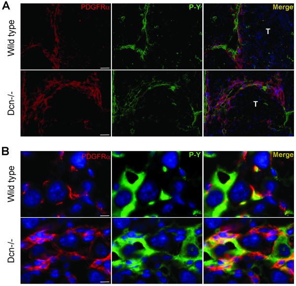
Fig. 5.
Localization of PDGFRα in TA-treated livers of wild type and decorin-null animals. (A): PDGFRα and phospho-tyrosine (P-Y) double immunostaining in cirrhotic septa of wild type (Wt) and decorin-null (Dcn−/−) liver sections. Scale bar=100 μm. (B): Tumor cells in wild type (1st row) and Dcn−/− (2nd row) TA-treated livers stained by anti-PDGFRα and phospho-tyrosine (P-Y) antibodies. Scale bar=10 μm.

