
Figure 2.
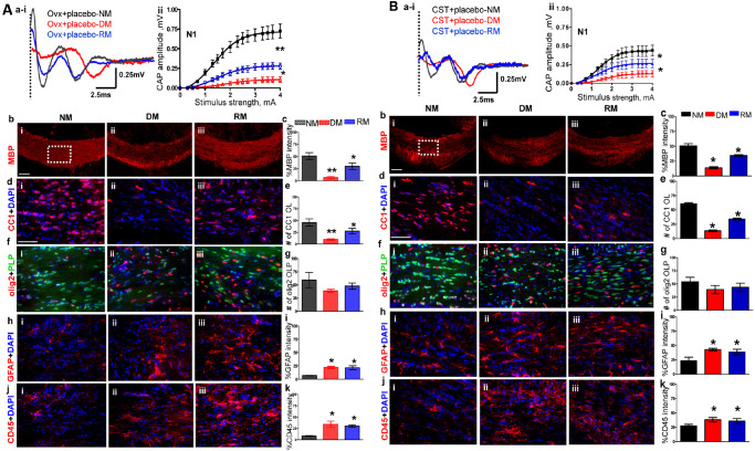
A. Ovariectomy (OVX) increases severity of demyelination and reduces efficiency of remyelination in females. (ai,ii) CAP responses for OVX females during NM (black), DM (red) and RM (blue). Representative traces for CAP response to 4.0 mA stimulus in OVX females induced a significant decrease in N1 peak amplitude during NM, DM and RM as compared to gonadally intact female mice. Quantification of N1 CAP amplitude shows a significant decrease in peak N1 CAP amplitude during DM with incomplete recovery during RM. DM is more severe, and RM is less efficient in OVX females compared with gonadally intact females. (bi–iii, c) MBP immunoreactivity imaged at 10× from the CC under NM, DM and RM condition showed a significant decrease during DM and a significant recovery during RM. (di–iii, e) Consecutive brain sections were immunostained and imaged at 40× magnification from various groups in an area similar to that of the dashed square in bi. OLs stained with CC1 (red) and counterstained for nuclei with DAPI (blue). Cell loss is significant during DM with partial but significant recovery during RM. (fi–iii, g) OLPs expressing PLP_EGFP and stained with olig2 (red) show nonsignificant trends of decrease during DM and recovery during RM. Cell counts are significantly lower than gonadally intact counterparts. (hi–iii, i) Reactive astrocytes stained with GFAP (red) and counterstained for nuclei with DAPI (blue) show a significant increase in intensity that persists in DM and RM. (ji–iii, k) Microglia stained with CD45 (red) and counterstained with DAPI (blue) show a significant increase in immunoreactivity that persists during DM and RM (*P < 0.05; n = 6). B. Castration increases the severity of demyelination and reduces efficiency of remyelination in males. (ai–iii) CAP responses for castrated (CSTed) males during NM (black), DM (red) and RM (blue). Representative traces for CAP response to 4.0 mA stimulus in CSTed mice induced a significant decrease in N1 peak amplitude during NM, DM and RM as compared to gonadally intact mice. Quantification of N1 CAP amplitude shows severe N1 signal loss during DM with incomplete recovery during RM. CSTed mice showed a more severe DM‐induced decrease and less efficient RM‐induced increase in N1 amplitude as compared to gonadally intact males. (bi–iii, c) MBP levels were assessed by intensity changes in MBP (red) immunoreactivity during NM, DM and RM. MBP immunoreactivity imaged at 10× from the CC under NM, DM and RM. Quantification of MBP intensity showed a significant decrease during DM in CST males, with significantly less efficient recovery during RM as compared to gonadally intact males. (di–iii, e) Consecutive brain sections were immunostained and imaged at 40× magnification from various groups in an area similar to that of the dashed square in bi. Mature OLs stained with CC1 (red) and counterstained with DAPI (blue) showed a significant loss of cells during DM with partial but significant recovery during RM. (fi–iii, g) OLPs stained with olig2 (red) and PLP_EGFP (green) cells show nonsignificant trends of decrease during DM and recovery during RM. (hi–iii, i) GFAP+ astrocytes (red) counterstained with DAPI (blue) show a significant increase in intensity that persists in DM and RM conditions. (ji–iii, k) CD45+ microglia (red) counterstained with DAPI (blue) showed a significant increase in fluorescence intensity during DM that persisted during RM. in CST + placebo males as compared to gonadally intact mice (*P < 0.05; n = 8).