
Figure 3.
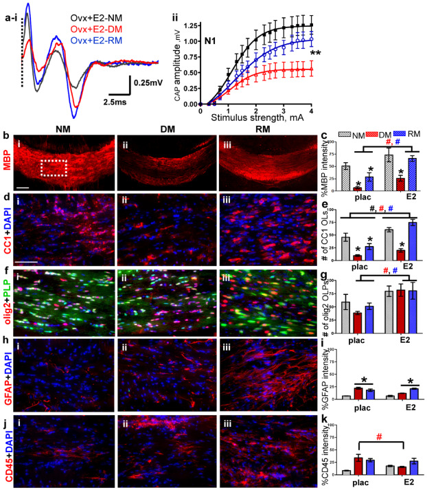
Estradiol (E2) hormone supplementation improves normal myelination, partially ameliorates demyelination and accelerates remyelination. (ai–ii) CAP responses for E2‐administered ovariectomized (OVX) females under NM (black), DM (red) and RM (blue) conditions. Representative traces for CAP response to 4.0 mA stimulus induced a significant increase in N1 peak amplitude during NM, DM and RM. Quantification of N1 CAP amplitudes reveals a significant increase in N1 peak amplitudes during NM, DM and RM as compared to placebo‐administered OVX and gonadally intact females. (bi–iii, c) Increased MBP (red) staining intensity during NM, DM and RM condition in OVX + E2 was observed as compared to OVX + placebo mice. (di–iii, e) Consecutive brain sections were immunostained for various markers and imaged at 40× magnification in an area similar to the one shown by the dashed squares in bi. Increased numbers of CC1+ OLs (red) counterstained with DAPI (blue) were present in CC of OVX + E2 mice compared to OVX + placebo mice. (fi–iii, g) Cells expressing PLP_EGFP and co‐stained with olig2 (red) had a sustained increase under NM, DM and RM condition as compared to OVX + E2 mice. (hi–iii, i) GFAP+ astrocytes (red) and counterstained with DAPI (blue) expressed increased fluorescence during DM and RM condition, but were similar to OVX + placebo group. (ji–iii, k) CD45+ (red) microglia counterstained with DAPI (blue) revealed similar levels of fluorescence intensity during DM and RM in OVX + E2 and OVX + placebo groups (*P < 0.05; n = 8–12); (ANOVA; #P < 0.5, n = 6–8; colors represent significance for NM, DM or RM).