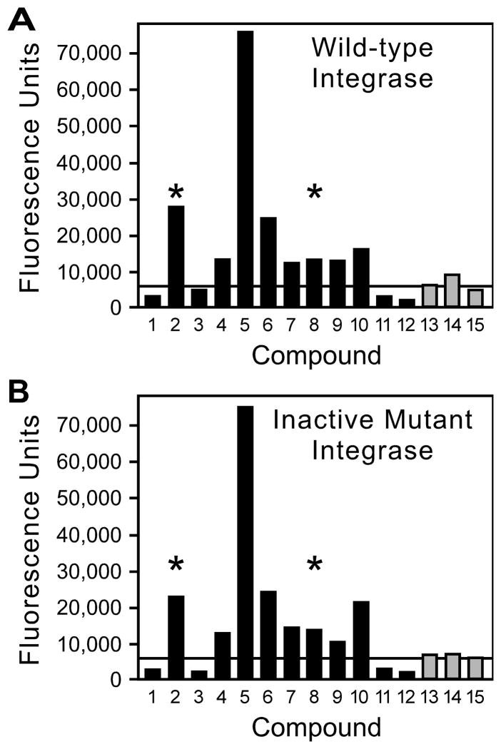
Figure 4. Examples of secondary assay.
Compounds that caused stimulation of signal in the primary screen were retested in parallel reactions that used wild-type integrase (panel A) or an active-site D116I mutant of integrase (panel B). Solid bars indicate compounds that had caused greatly increased fluorescence in the primary screen (reactions 1 to 12), and shaded bars denote compounds that had caused moderately increased fluorescence (reactions 13 to 15). Asterisks mark compounds for which visible color was noted in the chemical source plate or in the well of the secondary assay plate.

