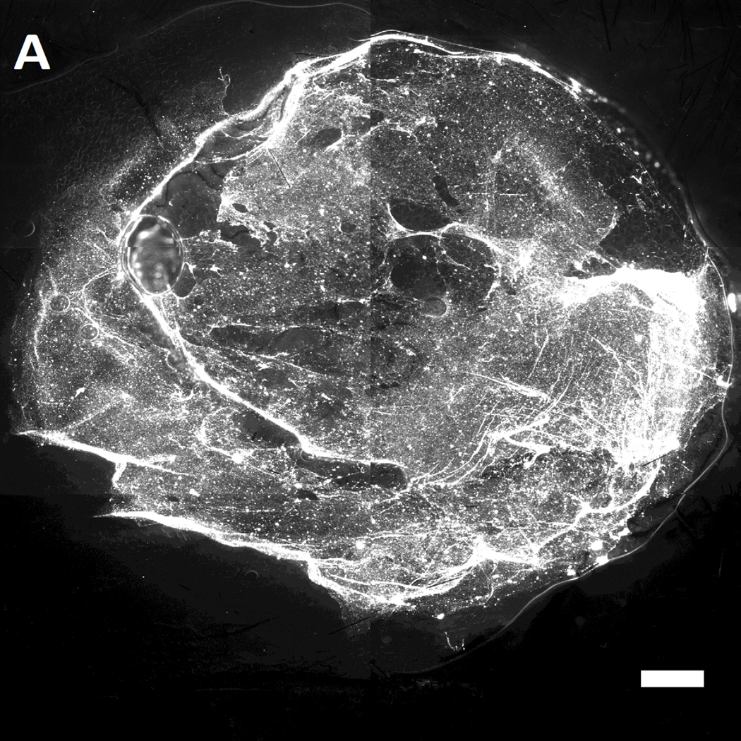
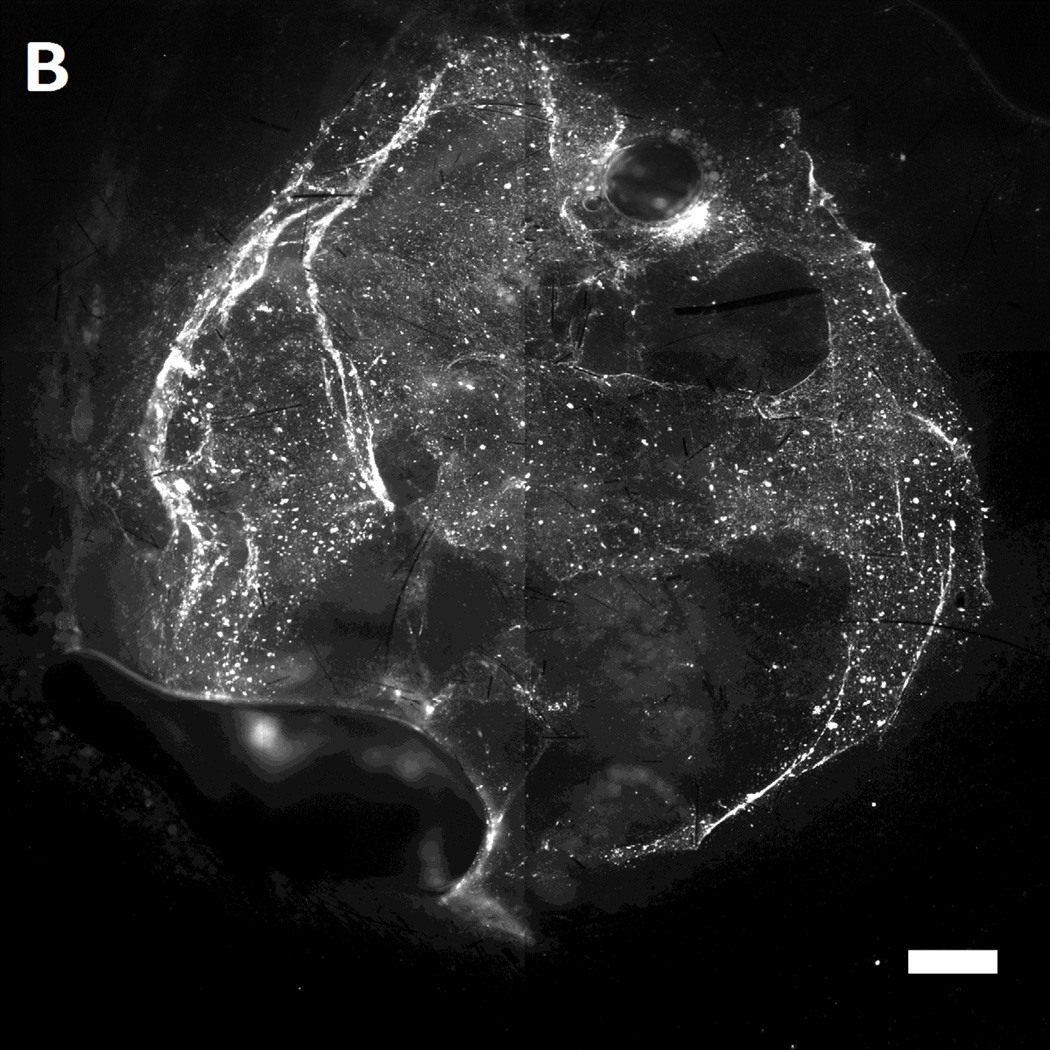
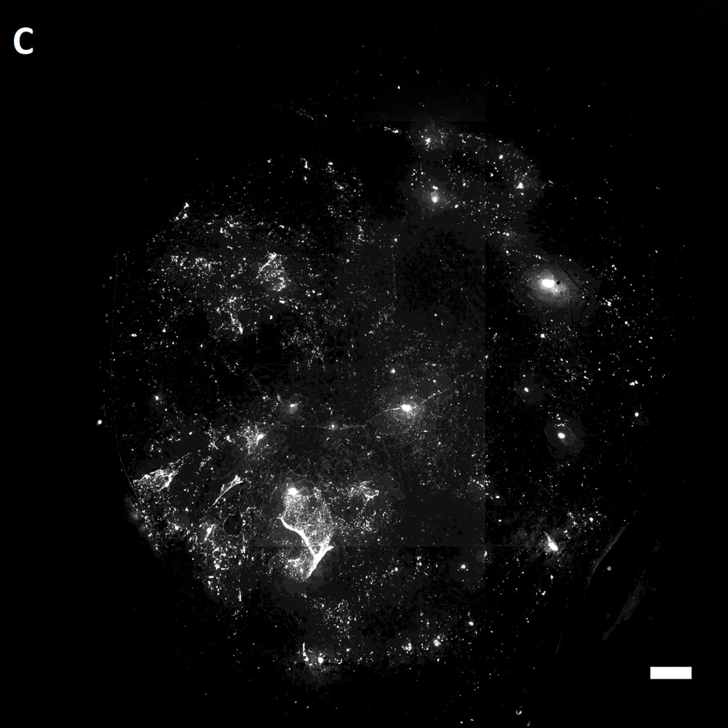
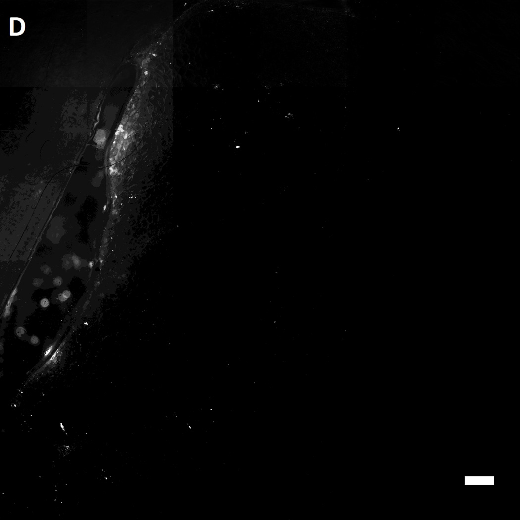
Figure 7.
Representative fluorescent images of live wild-type [A,B] and genetically diabetic [C,D] mice wounds (diameter: 4 mm.) treated with [A,C] sulfhydryl reactive microbeads (MAL-microbeads) and [B, D] non-reactive microbeads (NH2-microbeads) for 30 mins followed by rinse with PBS (pH 7.4). Scale bars: 500 µm


