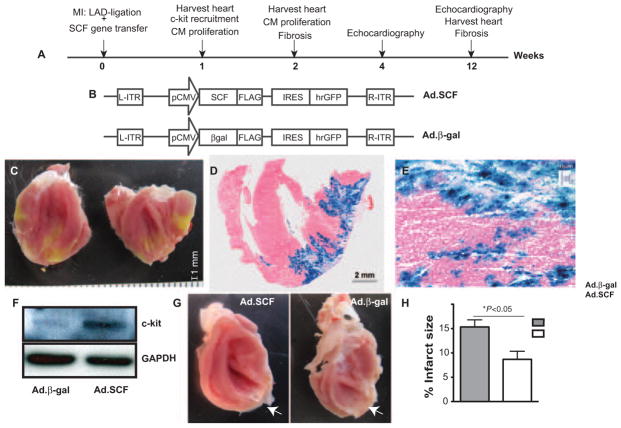
Figure 1. Study design.
A, Experimental timeline. Our strategy consisted of using stem cell factor (SCF) as a therapy in cardiac regeneration in rats undergoing myocardial infarction (MI) after delivering adenoviruses expressing SCF membrane-bound form and green fluorescent protein (Ad.SCF) or adenoviruses containing β-gal and green fluorescent protein (Ad.β-gal) recombinant adenoviruses in the peri-infarct area. Myocardial regeneration and function was assessed at 1, 2, 4, and 12 weeks post-MI. B, Recombinant Ad.SCF and Ad.β-gal adenoviruses were generated after cloning SCF membrane isoform and β-gal gene sequences, respectively, into an adenoviral plasmid containing GFP as a reporter gene. C–E, Detection of gene transfer within the myocardium 1 week post-MI. Distribution of the viral infection was confirmed by GFP expression, imaged through digital photography (C) and by X-gal staining (blue) (D and E) in control rats (Ad.β-gal). F, Analysis of c-kit receptor expression by western blot (WB), in lysates from control and Ad.SCF-treated hearts. α-GAPDH was used as protein loading control. G and H, Infarct size (white arrows) (G) was measured by cardiac magnetic resonance imaging 2 weeks post-MI (15.3±1.4% Ad.β-gal vs 8.6±1.6% Ad.SCF; *P<0.05; H). LAD indicates left anterior descending coronary artery; IRES, internal ribosomal entry site.

