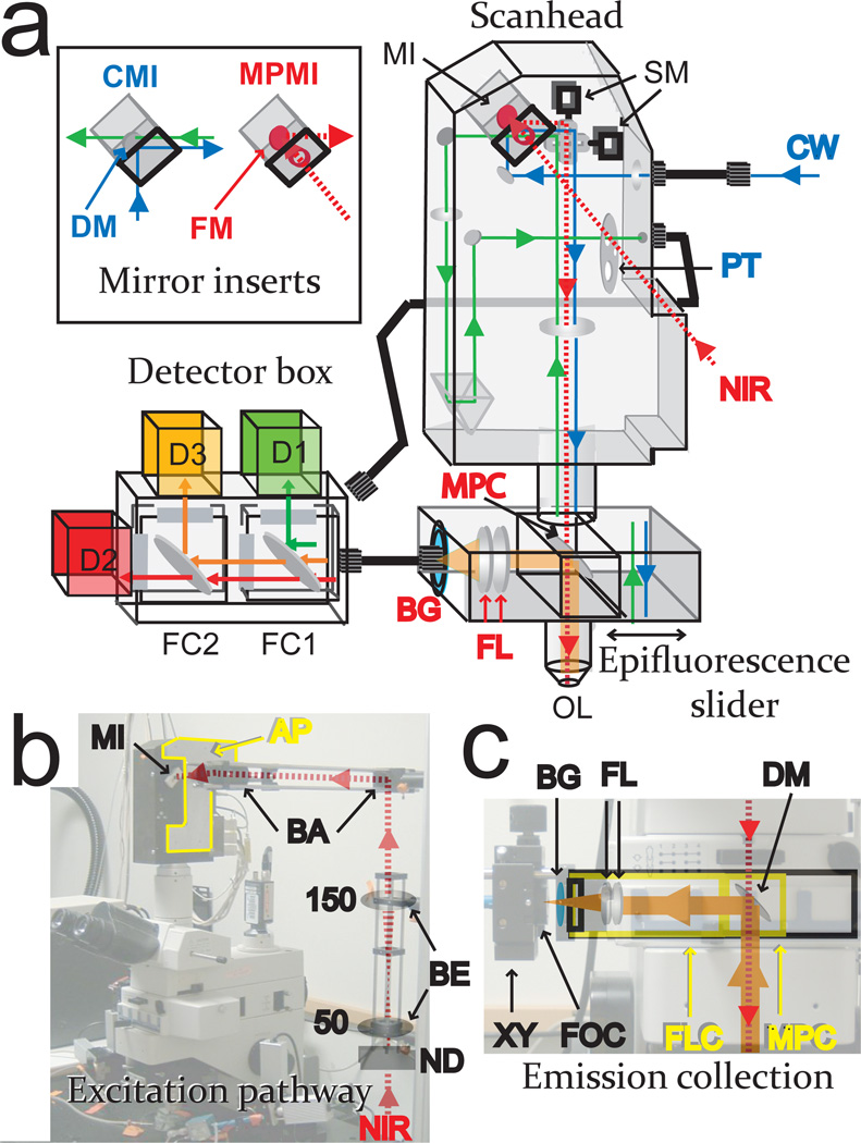
Figure 1.
Confocal to multiphoton conversion. (a) Schematic of system adaptation. The near infrared (NIR) excitation path is shown in red, multiphoton-excited emission in orange, CW excitation in blue, and standard single photon-excited emission in green. Components utilized in both configurations are labeled black, in confocal only blue, and in multiphoton only red. The setup is shown in the multiphoton configuration, where the detector unit is connected by multimode fiber optic cable to the epifluorescence slider and not the scanhead. Abbreviations: mirror insert (MI), scan mirrors (SM), pinhole turret (PT), multiphoton cube (MPC), focusing lens (FL), blue glass filter (BG), objective lens (OL). Inset: Confocal mirror insert (CMI) using dichroic mirror (DM), multiphoton mirror insert (MPMI) using full mirror (FM). Detector: emission filter cubes (FC1,2), and detectors (D1,2,3). (b) Multiphoton excitation pathway with NIR beam passing neutral density filter (ND), telescoping beam expander (BE) consisting of 50 and 150mm focal length achromats, and beam aligner (BA) before entering custom-made mirror insert (MI). Also shown is the custom-made adapter plate (AP) (highlighted in yellow) connecting the scan head to the external optics. (c) Multiphoton epi-fluorescence emission pathway. Two special cubes were connected to the rail inside the slider by means of its dovetail and clamping screws: A custom-made focusing cube (FLC, highlighted in yellow) was secured to the leftmost (as shown) part of the rail. This cube contains the 17.5mm focal length lens combination (FL) and the 25mm diameter tube that serves to anchor the microbench parts holding the BG39 emission filter, (BG), X–Y positioner (XY), and fiberoptic coupling (FOC). The multiphoton cube (MPC, highlighted in yellow) containing the dichroic mirror (DM) is oriented such that the collected fluorescence is directed away from the scanhead and towards the external detector unit, is then fastened to the same rail immediately adjacent to the focusing cube. The slider is placed in position 2 (as shown) for multiphoton microscopy and position 1 to remove the dichroic mirror from the beam path in confocal mode.

