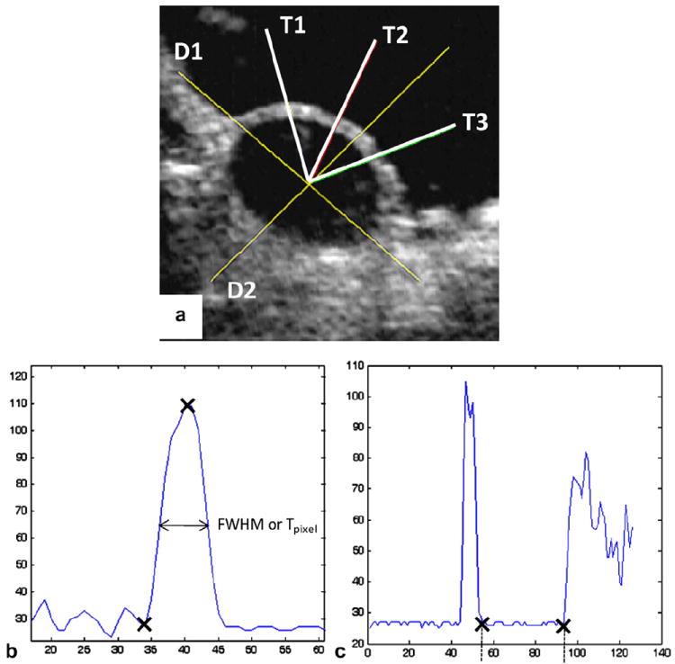
Fig. 2.

(a) An ultrasound image of a cross-section of the inflated CS vessel, with two line segments D1 and D2 for measuring diameters and three line segments T1, T2 and T3 for thickness measurements; (b) thickness measurements using the FWHM method (to obtain Tpixel, or the thickness in pixels, of the width of the half amplitude of the peak); (c) diameter measurements (distance between the two crosses, in pixels), both processed by the Matlab “improfile” function.
