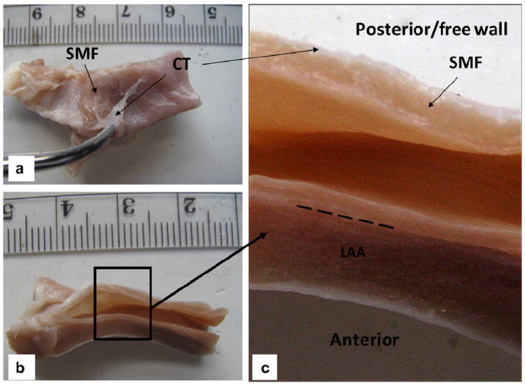
Fig. 5.

(a) Coronary sinus (CS) vessel fixed in 10% formalin for 2 days, with the connective tissue (CT) peeled off to expose the striated myocardial fiber (SMF) layer; (b) axial view; (c) a closer view of the CS inner wall with distinctive layers of SMF, CT and left atrial appendage (LAA). The long dashed line indicates the barrier between the CS wall and LAA.
