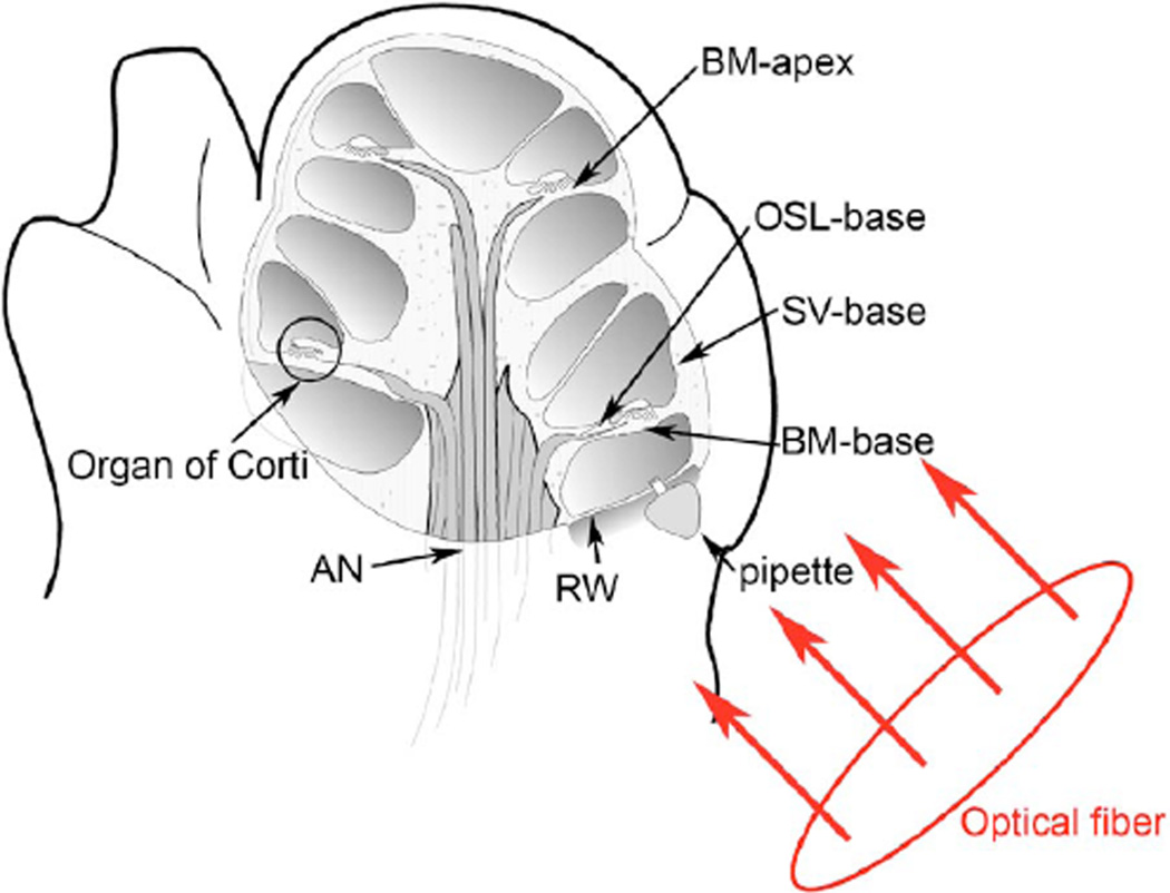
Fig. 1.
Diagram of the mouse cochlea with the orientation of the optical fiber. A midmodiolar cross section through the cochlea cuts perpendicular to the organ of Corti. The pipette used to perfuse trypan blue to stain the basilar membrane at the base of the cochlea (BM-base) was inserted through the round window membrane (RW): basilar membrane at the apex of the cochlea (BM-apex), osseous spiral lamina (OSL-base), stria vascularis (SV-base), and auditory nerve (AN).

