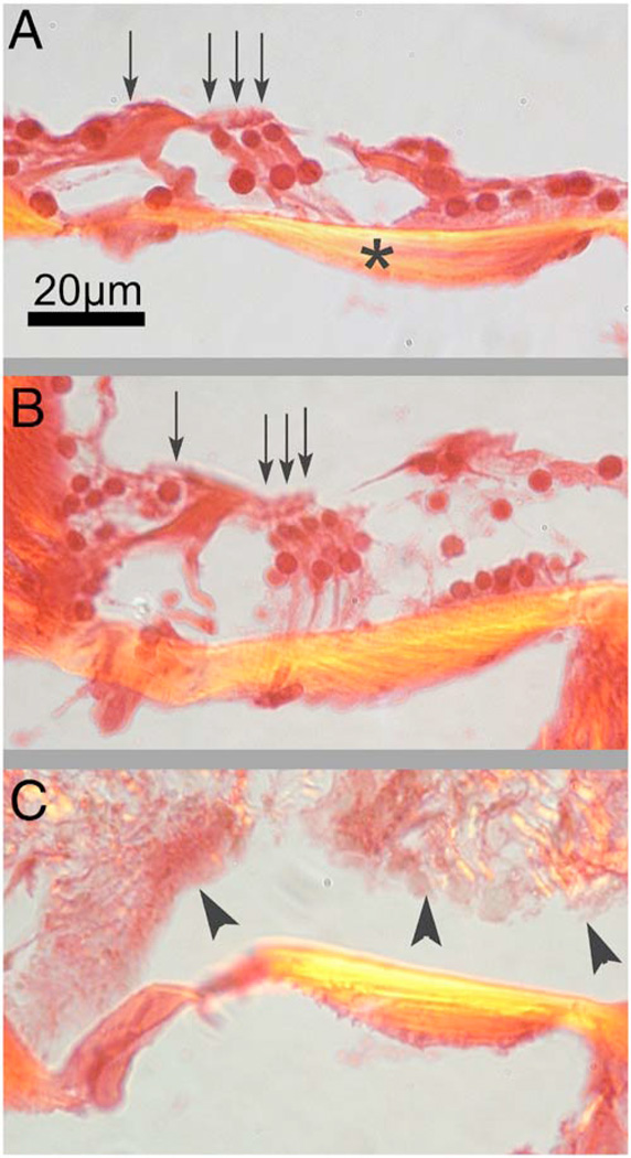
Fig. 2.
Organ of Corti. Paraffin-embedded cochlear cross sections stained with picrosirius red and hemotoxylin were visualized using polarization microscopy to highlight collagen arrays. (A) A representative control section (cochlea not perfused or irradiated). The normal basilar membrane birefringence can be noted (*). Inner and outer hair cells are also visible (arrows). (B) A representative section of a cochlea 14 days after the laser irradiation with 15 J /cm2. Note the continued presence of hair cells (arrows) and the normal architecture of the organ of Corti. (C) A representative section of a cochlea 14 days after irradiation with 180 J /cm2. The basilar membrane birefringence appears brighter (more yellow) than that of the control cochlea. Hair cells are no longer present and scar tissue fills the intracochlear scalae (arrowheads).

