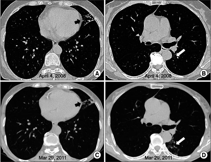
Figure 1.
A 69-year-old woman with bronchiectasis and nontuberculous mycobacterial lung disease caused by Mycobacterium chelonae. (A) A transverse computed tomography (CT) scan (2.5-mm-section thickness) at the time of presentation reveals bronchiectasis and bronchiolitis in the lingular division of the left upper lobe. (B) Mild bronchiectasis was noted in the superior segment of the left lower lobe at the time of presentation. (C) A CT scan of the same patient at 3 years after diagnosis reveals mild progression of peribronchial infiltration on the lingular division of the left upper lobe. (D) There was newly appearing bronchiolitis in the superior segment of the left lower lobe.

