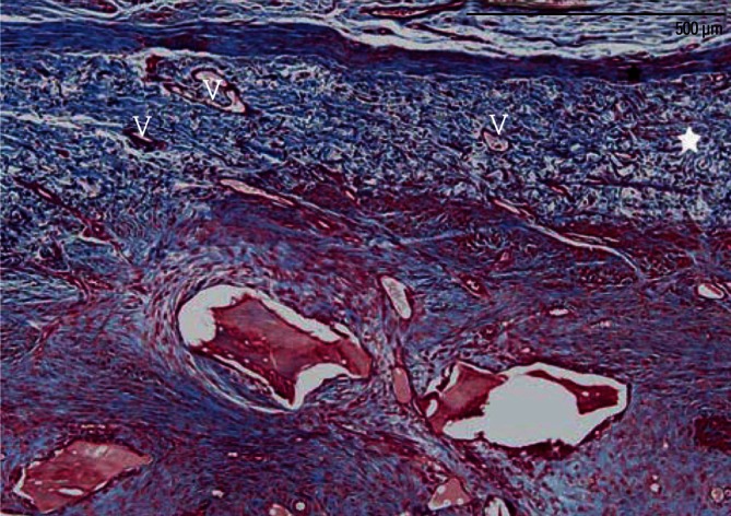
Figure 6.

A high-magnification photomicrograph of a graft site of the membrane-cover group, showing many newly formed vessels (V) within the residual collagen membrane (white asterisk). Black asterisk: periosteum-like dense connective tissue layer (Masson's trichrome stain, scale bar=500 µm).
