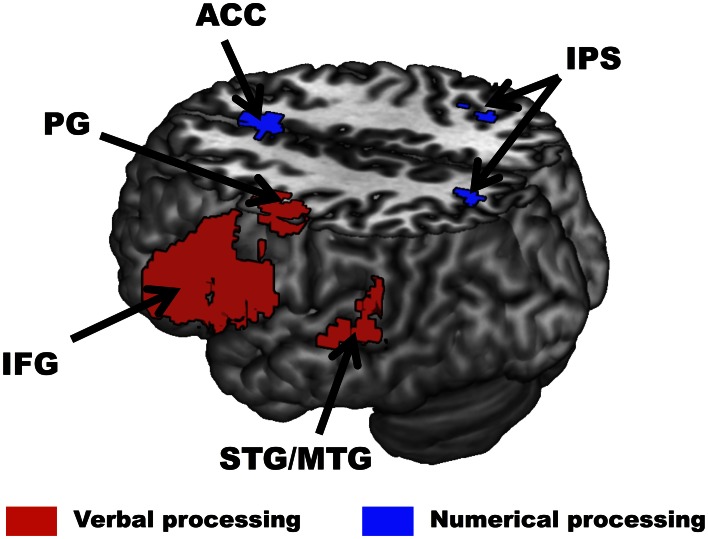
Figure 3.
Brain regions identified by the localizer scans across all participants. In the verbal processing localizer (red), greater activity for words than symbols was observed in the left Superior and Middle Temporal Gyri (STG/MTG), left Inferior Frontal Gyrus (IFG), and left Precentral Gyrus (PG). In the numerical processing localizer (blue), greater activity for difficult than easy comparisons was observed in the right Intraparietal Sulcus (IPS) and Anterior Cingulate Cortex (ACC). All activations are overlaid on a 3D rendering of the MNI-normalized anatomical brain. The upper part of the brain is cut out (Z = 46) to show activations in deeper sulci and along the medial wall of the cortex.

