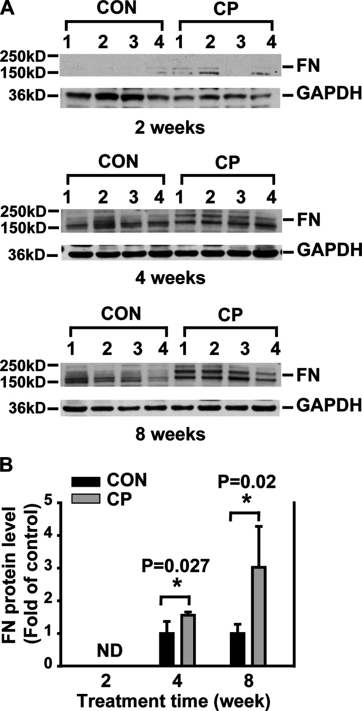
Fig. 2.
Time-dependent increase of fibronectin (FN) in CP mice. A: Western blotting of pancreas tissue lysates from CON and CP mice with anti-FN antibody. B: quantification of the Western blots. The protein levels were normalized against GAPDH and quantified as fold of control. ND, nondetectable. *P < 0.05 compared with control; n = 4 mice/group.

