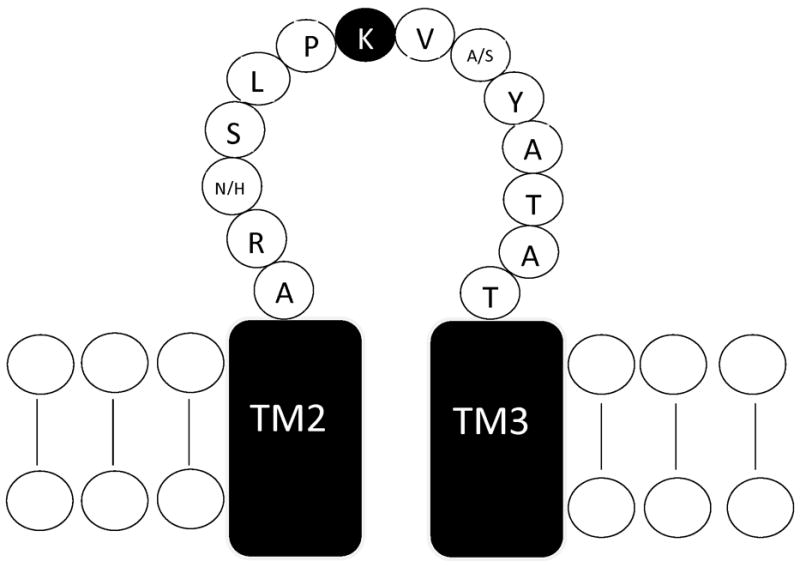
Figure 1. Location of mutation site.

Schematic representation of the TM2–TM3 extracellular domain of the GABAAR subunit. The conserved lysine residue mutated in this study is indicated by the filled circle. The α1 and α6 subunits differ at only two residues within in this domain, shown as α1/α6. Sequence from [17].
